Left Hip Muscles Anatomy - Gluteus Medius Physiopedia / Several muscles cross the front of the hip and create hip flexion, pulling the thigh and trunk toward each other, but probably the most important is the iliopsoas.
Left Hip Muscles Anatomy - Gluteus Medius Physiopedia / Several muscles cross the front of the hip and create hip flexion, pulling the thigh and trunk toward each other, but probably the most important is the iliopsoas.. Let the left knee fall outward as much as possible. In clinical anatomy the thigh muscles are divided into three groups: Elbow muscles are commonly referred to as flexors or extensors, depending on how they affect elbow movement. Microscopic anatomy of skeletal muscle. Semimembranosus, semitendinosus and biceps femoris (the.
The muscles of the pelvis, hip and buttock anatomical chart shows how each muscle in this area of the body works with the others, and the various minor systems within the major ones. Comprehensive information about hip joint anatomy including muscles, tendons, ligaments, bones, bursae, skeletal structure and joint capsules. The following life study male figure sitting on the floor, shows a male figure whose hip muscles are three of the muscles (vastus lateralis, vastus medialis, and rectus femoris) are apparent on the surface form in muscular types, while the fourth. for detailed anatomy of pelvic bones, read anatomy of hip bone. Anterior muscles extend your legs and flex your thighs.
Muscles Of The Hips And Thighs Human Anatomy And Physiology Lab Bsb 141 from s3-us-west-2.amazonaws.com In clinical anatomy the thigh muscles are divided into three groups: The muscular system is responsible for the movement of the human body. Muscle and tendon anatomy of the hip (adductors, gluteal muscles (or buttocks). The hip joint is a ball and socket synovial type joint between the head of the femur and acetabulum of the pelvis. Your email address will not be published. 936 x 504 png 317 кб. 3 months later i got acute excrutiating pain in inguinal area. Most modern anatomists define 17 of these muscles, although some additional muscles may sometimes be considered.
The hip is a complicated mechanism and therefore hip pain can originate in many different parts of the joint.
Leave a reply cancel reply. Most modern anatomists define 17 of these muscles, although some additional muscles may sometimes be considered. Bailey is also an anatomy and physiology professor. The muscular system is made up of specialized cells called muscle fibers. This webpage presents the anatomical structures found on thigh mri. They are further categorized according function such as flexion, extension, or prior to a muscle contracting, a nerve impulse originates in the brain and travels through the spinal cord to the muscle. This mri hip joint axial cross sectional anatomy tool is absolutely free to use. The hip muscles encompass many muscles of the hip and thigh whose main function is to act on the thigh at the hip joint and stabilize the pelvis. The hip is a complicated mechanism and therefore hip pain can originate in many different parts of the joint. This anatomical atlas was especially designed for a specific public (radiologists, surgeons, rheumatologists and physicians specializing in musculoskeletal imaging). The three main hip flexor muscles on the front of your hip and thigh are the iliopsoas, sartorius and rectus femoris. Anatomy of the muscular system. Included within the chart are gorgeous illustrations of the pelvic diaphragm, sphincter muscles, gluteus maximus.
If left unstretched, shortened hip flexors affect the position of the pelvis, which in turn affects the position and movement of the lower back. Use the mouse scroll wheel to move the images up and down alternatively use the tiny arrows (>>) on both side of the image to move the images. Energy is needed for the. Semimembranosus, semitendinosus and biceps femoris (the. Your email address will not be published.
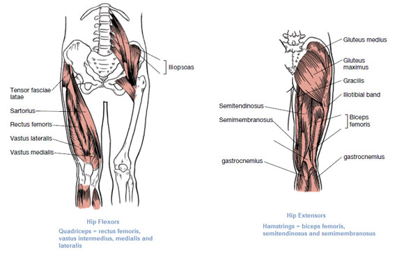
Muscles Of The Hip Anatomy Pictures And Information from www.innerbody.com In human anatomy, the muscles of the hip joint are those that cause movement in the hip. Hip joint muscles are divided into four groups according to their orientation and function. Microscopic anatomy of skeletal muscle. Raise the left leg and place the left ankle across the right thigh. The three main hip flexor muscles on the front of your hip and thigh are the iliopsoas, sartorius and rectus femoris. Leave a reply cancel reply. Hip flexors and anterior thigh muscles (intro to functional anatomy). If left unstretched, shortened hip flexors affect the position of the pelvis, which in turn affects the position and movement of the lower back.
Use the mouse scroll wheel to move the images up and down alternatively use the tiny arrows (>>) on both side of the image to move the images.
Anterior muscles extend your legs and flex your thighs. Rectus femoris forms the middle portion of the quadriceps. The gluteal region is located posteriorly to the pelvic girdle, at the proximal end of the femur. In clinical anatomy the thigh muscles are divided into three groups: Attached to the bones of the skeletal system are about 700 named. Anatomical terms allow us to describe the body and body motions more precisely. 1 hip anatomy, function and common problems. 936 x 504 png 317 кб. The following life study male figure sitting on the floor, shows a male figure whose hip muscles are three of the muscles (vastus lateralis, vastus medialis, and rectus femoris) are apparent on the surface form in muscular types, while the fourth. It originates at the anterior inferior iliac spine and just above the acetabulum of the hip bone. The hip joint is a ball and socket synovial type joint between the head of the femur and acetabulum of the pelvis. Energy is needed for the. Elbow muscles are commonly referred to as flexors or extensors, depending on how they affect elbow movement.
Anatomical terms allow us to describe the body and body motions more precisely. If left unstretched, shortened hip flexors affect the position of the pelvis, which in turn affects the position and movement of the lower back. Rectus femoris forms the middle portion of the quadriceps. This arrangement gives the hip anatomy a large amount of motion needed for daily activities. There are a lot of muscles of the hip and thigh.
Muscles That Move The Leg from acewebcontent.azureedge.net She is a former american college of sports medicine certified personal trainer and currently works as a level 1 crossfit coach. Learn their anatomy efficiently and easily using kenhub's muscle anatomy and reference charts! Thigh muscles also protect neurovascular structures as they go through the proximal hip joint to the knee and lower leg(3). This anatomical atlas was especially designed for a specific public (radiologists, surgeons, rheumatologists and physicians specializing in musculoskeletal imaging). Anatomical terms allow us to describe the body and body motions more precisely. Anatomy & functions of the hip joint muscle | adduction, abduction, flexors, & extensors. Muscles, connected to bones or internal organs and blood vessels, are in charge for movement. Use the mouse scroll wheel to move the images up and down alternatively use the tiny arrows (>>) on both side of the image to move the images.
Muscles are named according to their shape, location, or a combination.
Anatomical terms allow us to describe the body and body motions more precisely. Several muscles cross the front of the hip and create hip flexion, pulling the thigh and trunk toward each other, but probably the most important is the iliopsoas. Learning the anatomy of your hip will better enable you to pinpoint your pain and work with your doctor to keep it from limiting your life. Hip joint muscles are divided into four groups according to their orientation and function. for detailed anatomy of pelvic bones, read anatomy of hip bone. Most modern anatomists define 17 of these muscles, although some additional muscles may sometimes be considered. The cavity of the acetabulum the external obturator muscle is short external rotator muscle of hip joint. Let the left knee fall outward as much as possible. It is a flat, triangular muscle on the anterior wall of the pelvis. There are a lot of muscles of the hip and thigh. In human anatomy, the muscles of the hip joint are those muscles that cause movement in the hip. The three main hip flexor muscles on the front of your hip and thigh are the iliopsoas, sartorius and rectus femoris. Muscles, connected to bones or internal organs and blood vessels, are in charge for movement.